Real Cloacal Exstrophy Pictures: Diagnosis, Anatomy & Treatment Overview
Whether you are the parent of a child with Cloacal Exstrophy, a medical student, or someone for whom it is a new birth condition, the reality of seeing actual photos or pictures of Cloacal Exstrophy can be overwhelming. Since the illness impacts multiple organs and bodies systems it is highly likely that the images will appear medically complex, hard to process emotionally. But knowing what the images portray can help to educate people about the condition, how it will be diagnosed and the treatment journey thereafter.
Cloacal exstrophy is a very rare and severe form of abdominal wall birth defect. It is present from birth, and develops in the bladder, intestinal tract, sexual organs, pelvic bones and lower portion of the abdominal wall. Surgery may be necessary soon after birth in many instances, followed by medical treatment throughout childhood.
This informative article will seek to describe the features that are usually observed in cloacal exstrophy photos, how the condition is diagnosed, cloacal exstrophy anatomy, cloacal exstrophy treatments and what can be expected for the future for the family. To deliver information that is clear, useful, respectful and comprehensible.
The Question is what is cloacal exstrophy?
Cloacal exstrophy is a rare congenital disorder which is present at birth. Occurs very early in fetal development when portions of the lower abdominal wall don’t develop properly.
It is part of a larger family of abnormalities, called the OEIS complex. OEIS stands for:
- Omphalocele
- Bladder outside and up
- Imperforate anus
- Spinal defects
Many children have abnormalities with several of these features, not every child has all of these abnormalities, but many have several.
In cloacal exstrophy, the bladder is split open and exposed outside the body. Some of the intestine may also protrude from the abdominal wall. The genital area may be different and the pelvic bones may be farther apart than normal.
This condition involves a number of different systems, so a team of more than one doctor may be involved, such as the orthopedic surgeons, urologists, neonatal care specialists and pediatric surgeons.
What does real cloacal exstrophy pictures indicate?
Typically, images of the cloacal exstrophy are not obtained for aesthetic reasons but rather for learning or diagnosis and perhaps for surgery-planning purposes. These photographs will depend on the severity of the disorder and/or the age of the child.
When taking photographs of newborns, a number of things can be observed:
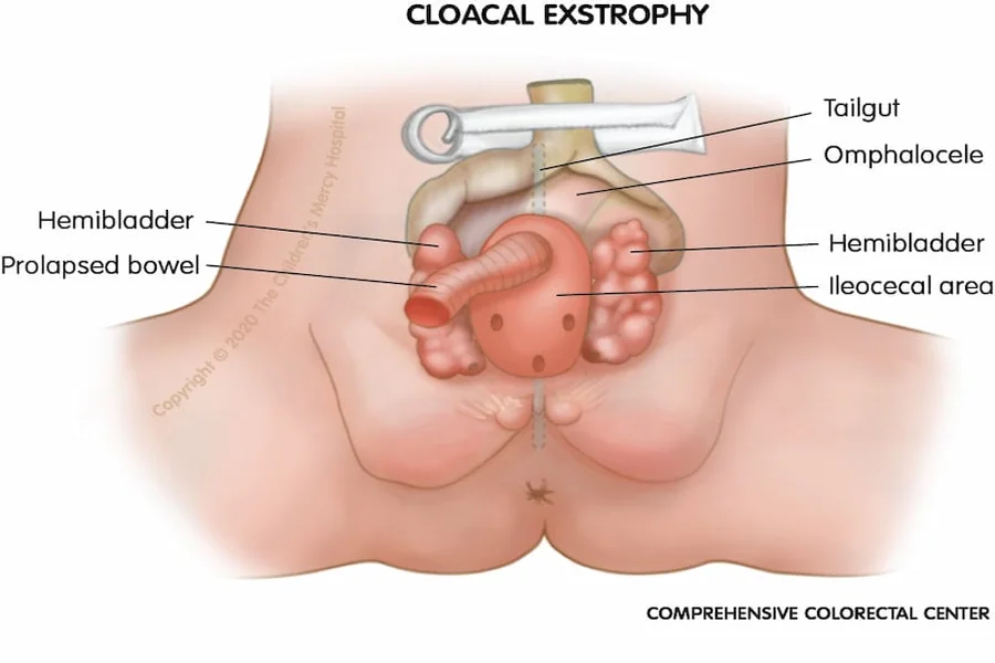
The lower part of the abdominal wall may be open and tissue may be exposed in the center of the abdomen. Bladder may be split lengthwise in half. In between these halves a portion of the intestine may be found poking out.
A few pictures also demonstrate an omphalocele, a sac of belly organs that bulge outward around the umbilicus or belly button.
The genital area may appear retarded or split. Penis can be shortened or split in boys. In female, the clitoris may be divided into two parts. The anus may be defective or seem to have been missing.
Radiograms or PICs of the insides might reveal enlarged pelvic bones or abnormalities in the spine.
Keep in mind that these images are not for decoration but are medical images. They assist doctors in understanding anatomy, planning surgery and monitoring treatment process. They should always be looked at in a respectful and healthcare setting.
Explain to the patient why they should be concerned about the condition, including the bodily and physiological effects.
For purposes of understanding cloacal exstrophy, it is important to realize how normal fetal development occurs.
As the pregnancy advances, the lower abdominal wall and internal organs start to split & close up accordingly. In cloacal exstrophy this developmental process is disturbed.
As a result:
Normally the bladder doesn’t close the way it should.
The intestines may remain exposed
The pelvic bones might not fuse completely
Reproductive organs can have varying development.
The anal opening might not develop properly.
Since there are a number of structures involved all at once, it would appear very complicated in medical images.
However, a misconception would be to think that every child with cloacal exstrophy is exactly the same. In practice, there is a continuum of the condition. Some babies have milder forms and some are more severely affected with abnormalities of the spine, kidneys or limbs.
Imaging tests, including ultrasound, MRI and X-rays, are typically used to get a complete understanding of how things are inside to plan surgery.
How Cloacal Exstrophy Is Diagnosed
Many times cloacal exstrophy is diagnosed before birth during a prenatal ultrasound.
Doctors might find indications like:
No bladder or abnormal bladder
Defects of the lower abdominal wall.
Spinal abnormalities
Omphalocele
Abnormal orientation of pelvic bones
Fetal MRI (magnetic resonance imaging) is sometimes used in order to get more detailed images. Prenatal diagnosis allows doctors to plan for the delivery and newborn treatment.
The diagnosis is usually confirmed by physical examination and imaging tests after birth. As it can be easily seen in most infants, the physician will probably be able to detect it rapidly.
Further testing may be performed such as:
Kidney ultrasound
Spinal imaging
Echocardiogram to check the heart
Tests of blood and urine.
Diagnosis at an early age is very critical as a baby with cloacal exstrophy may require special medical attention shortly after birth.
Therapy options and surgical care.
The treatment for cloacal exstrophy is very personal. No one surgery cures all. Rather, treatment commonly occurs in phases over an extended period—often for many years.
It may be necessary for initial surgeries after the baby is born (I.S.A.B).
Surgeons immediately after birth concentrate on covering organs which are exposed and stabilizing the baby’s state.
The initial surgery could include:
Closure of the abdominal wall in the absence of scar tissue.
Reconstructing the bladder
Segmentation & relocation of the gut.
Formation of colostomy as needed
In a colostomy the stool leaves the body through an opening in the abdomen while reconstructive surgery can be done at a later stage.
Reconstructive Surgery for the Bladder
Surgeries later in the child’s life can sometimes further enhance bladder function and urinary control.
Some children are able to gain some control over their bladders while others may need to access the bladder with a catheter or urinary diversion procedures from time to time.
Kidney function is monitored closely throughout life; urinary problems can make kid infection or injury more likely.
Orthopedic/Pelvic Surgery
Occasionally, the pelvic bones are widely spread apart and orthopedic surgery can aid in stabilizing the pelvis and improving mobility.
Several children will also need to be treated for spinal problems related to OEIS complex.
Genital Reconstruction
Later in childhood or at adolescence, reconstructive surgery of genital differences might take place. Plans for treatment and care are tailored to a person’s anatomy, function and family goals.
New surgical procedures still enhance the long-term prognosis of many surgical patients.
It is now time to consider the possibility of life after treatment, or long-term outcome.
Even though children are diagnosed with cloacal exstrophy, they typically travel a long path marked with doctors, but a lot of them lead active and fulfilling lives.
Long term care can include:
Ongoing surgeries
Physical therapy
Urology follow-up
Bowel management programs
Psychological support
Emotions and stress in the family can be significant at the beginning, after diagnosis. Building access to the existing healthcare facilities and support groups can have a big impact.
An important point is that survival and quality of life has increased greatly over the past few decades. Thanks to the developments in neonatal therapy, paediatric surgery, reconstructive medicine, many children are now able to reach adulthood.
But difficulties may be encountered. Long term management may include urinary tract infections, stooling problems, and frequent surgeries.
Parent/parents are often asked or encouraged to work on emotional development, education and social support of the child as well as medical treatment.
Common Misunderstandings About Cloacal Exstrophy Pictures
When medical pictures are on the internet sometimes they can be confusing or fearful when seen without a proper explanation.
A common error is thinking that photographs represent permanent anatomy that has not been treated. Actually, numerous pictures are snapped right after birth, before corrective surgery starts.
Another misconception is that all patients will achieve the same longer term results. Treatment success is highly unpredictable for reasons of anatomy, related conditions and access to specialized care.
Also, images on the web may not be properly medically set. When a child or young person has a rarely talked about health condition, it’s always best to go to trusted medical professionals, paediatric wards or educational health providers for information.
Parents must not act on the basis of pictures they see on the internet and self diagnose. Imaging findings should never be interpreted alone and without the guidance of a healthcare professional, who will be able to guide an individual.
Read Also: Questions to Ask Before Choosing a Sperm Donor
Final Thoughts
Cloacal Exstrophy is a rare and medically complex condition which impacts several areas of the body all at once. Education with real medical photos, particularly, can be helpful for health care providers, medical students, and families to glean information about the anatomy – but should be done with sensitivity and proper medical explanation.
Diagnosis of this condition can be scary, but with today’s multidisciplinary care and pediatric surgery, treatment options have become very successful. Most of the children who have reconstructive surgery live normal lives and continue to grow and learn.
The most crucial thing for parents to do when they receive this diagnosis is to make a connection with experts with great experience in their field who are well aware of the diagnosis. Knowledge, preparation, and long-term support can make the journey more manageable and hopeful.
FAQs
Is cloacal exstrophy very rare?
Yes. Cloacal exstrophy is extremely rare and occurs in only a small number of births worldwide. Because of its rarity, treatment is usually handled at specialized pediatric centers.
Can cloacal exstrophy be detected during pregnancy?
In many cases, yes. Prenatal ultrasound and fetal MRI can often identify signs of the condition before birth.
Do all babies with cloacal exstrophy need surgery?
Yes. Surgical treatment is usually necessary soon after birth to protect organs and reconstruct affected body structures.
Can children with cloacal exstrophy live normal lives?
Many children grow into adulthood and lead productive lives, although they may require ongoing medical care and multiple surgeries over time.
Why are medical pictures of cloacal exstrophy important?
Pictures help doctors study anatomy, diagnose the condition, plan surgeries, and educate healthcare professionals. They can also help families better understand the condition when explained properly.
Is cloacal exstrophy the same as bladder exstrophy?
No. Cloacal exstrophy is generally more severe and involves additional organs such as the intestines and reproductive structures, while bladder exstrophy mainly affects the bladder and lower abdominal wall.